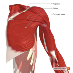
Shoulder Tendon Anatomy : They all attach to the greater tuberosity.. Around the shoulder, muscles in the back, neck, shoulder, chest and upper arm all work together to support and move the shoulder. About eight shoulder muscles attach to the. The long head of biceps (lhb) is a very important tendon that travels through the shoulder joint (glenohumeral joint). Muscles in turn move bones by pulling on the tendons. A tendon is a structure that connects muscle to bone, and the biceps are connected by tendons at both the elbow and shoulder joints.
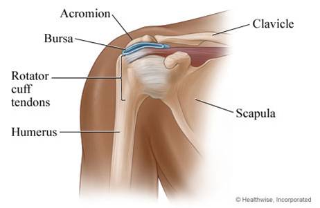
Your injury may range from mild inflammation to severe inflammation of most of your rotator cuff. The upper portion of the bicep also has a tendon that attaches it to the bones within the shoulder. The subacromial bursa reduces friction beneath the deltoid, promoting free motion of the rotator cuff tendons. Sichere dir kletterzubehör von tendon beim outdoor experten! Your rotator cuff consists of the muscles and tendons in your shoulder.
Shoulder Physiopedia from www.physio-pedia.com The muscle belly then crosses the entire upper arm and separates into two tendons. Shoulder anatomy joint isolated on white clipping path. 17 photos of the diagram of shoulder muscles and tendons. Shoulder mri assesses the following tendon and muscle structures: The biceps muscle has two tendons at the shoulder, called the long head and short head. Rotator cuff tears, biceps tendon tear at the shoulder. The bursa is a small sac of fluid that cushions and. A large humeral head lying on an almost flat scapular surface.
Tendons are much like ligaments, except that tendons attach muscles to bones.
Impingement syndrome is a condition where the rotator cuff tendons get pinched as they pass between the upper arm and tip of the shoulder. The upper portion of the bicep also has a tendon that attaches it to the bones within the shoulder. The muscles of the rotator cuff keep the humerus tightly in. It reduces wear and tear on the tendon during movement at the shoulder joint. Shoulder tendons chart ~ labeled anatomy chart of shoulder ligaments on white background stocktrek images. The tendons involved in the shoulder mainly include the long head of the biceps tendon and the tendons of the rotator cuff: As a consequence the shoulder joint is highly mobile, where stability takes second place to mobility, as is evident from the shape of the osseous structures: Bones in shoulder, ligaments of the shoulder joint, parts of the shoulder joint, shoulder anatomy, shoulder joints and muscles, shoulder structure anatomy, shoulder tendon anatomy, shoulder tendons ligaments, human muscles, bones in shoulder, ligaments of the shoulder joint, parts of. The muscle belly then crosses the entire upper arm and separates into two tendons. The long head of biceps (lhb) is a very important tendon that travels through the shoulder joint (glenohumeral joint). This socket is called the glenoid. Together they assist in stabilizing the shoulder joint as well as in performing various arm. They connect your upper arm bone to your shoulder blade.
Other supporting tendons include the pectoralis minor, coracobrachialis and the short head of the biceps. The rotator cuff is important in many routine activities, and when injured can cause severe pain. The bursa is a small sac of fluid that cushions and. It is an important cause of anterior shoulder pain and it is usually seen in association with other shoulder pathologies, such as rotator cuff tears and shoulder impingement. The rotator cuff is a collection of muscles and tendons that surround the shoulder, giving it support and allowing a wide range of motion.
Shoulder Cartilage And Tendon Injuries My Doctor Online from mydoctor.kaiserpermanente.org Together they assist in stabilizing the shoulder joint as well as in performing various arm. The purpose of this atlas is to focus the reader's attention on a series of bone, ligament, muscle and tendon. The anatomy of the shoulder. Tendons have lower blood flow than muscle tissue and are therefore more. The rotator cuff is made of the tendons of subscapularis, supraspinatus, infraspinatus and teres minor muscle. Shoulder anatomy joint isolated on white clipping path. Posterior graphic of the shoulder. The shoulder joint is composed of the glenoid (the shallow shoulder socket) and the head of the upper arm bone known as the humerus (the ball).
The tendons involved in the shoulder mainly include the long head of the biceps tendon and the tendons of the rotator cuff:
It reduces wear and tear on the tendon during movement at the shoulder joint. As a consequence the shoulder joint is highly mobile, where stability takes second place to mobility, as is evident from the shape of the osseous structures: The rotator cuff is important in many routine activities, and when injured can cause severe pain. The muscles of the shoulder have a wide range of functions, including abduction, adduction, flexion, extension, internal and external rotation. The biceps muscle has tendons on each end of the muscle. At the shoulder, the two tendons both attach to the large flat bone in the upper trunk called the scapula. The tendons are the attachment of the muscle to the bone. Each of these muscles has its own tendons that support the humerus. 1 the central bony structure of the shoulder is the scapula, where all of the muscles interact. A muscle contracts to move bones; Plastic study model of shoulder anatomy joint isolated on white background, clipping path. Shoulder anatomy joint isolated on white clipping path. The bursa is a small sac of fluid that cushions and.
Muscles move the bones by pulling on the tendons. Rotator cuff tears, biceps tendon tear at the shoulder. The shoulder joint is composed of the glenoid (the shallow shoulder socket) and the head of the upper arm bone known as the humerus (the ball). Rotator cuff and biceps tendon injuries are among the most common of these injuries. The biceps tendon runs from the biceps muscle, across the front of the shoulder, to the glenoid.
As The Shoulder Turns Understanding The Subscapularis Part I from www.sportsinjurybulletin.com The biceps muscle is located at the front of your upper arm. The head of your upper arm bone fits into a rounded socket in your shoulder blade. Other supporting tendons include the pectoralis minor, coracobrachialis and the short head of the biceps. Tendonitis of your shoulder is an inflammation of your rotator cuff or biceps tendon. The subacromial bursa reduces friction beneath the deltoid, promoting free motion of the rotator cuff tendons. The tendons are the attachment of the muscle to the bone. Tendons are much like ligaments, except that tendons attach muscles to bones. They connect your upper arm bone to your shoulder blade.
Shoulder mri assesses the following tendon and muscle structures:
Tendonitis of your shoulder is an inflammation of your rotator cuff or biceps tendon. The rotator cuff is made of the tendons of subscapularis, supraspinatus, infraspinatus and teres minor muscle. Muscles move the bones by pulling on the tendons. The biceps tendon runs from the biceps muscle, across the front of the shoulder, to the glenoid. A large humeral head lying on an almost flat scapular surface. The shoulder joint is composed of the glenoid (the shallow shoulder socket) and the head of the upper arm bone known as the humerus (the ball). The rotator cuff is a collection of muscles and tendons that surround the shoulder, giving it support and allowing a wide range of motion. One tendons inserts onto the forearm bone, the radius, and the second spreads out to join the. The bursa is a small sac of fluid that cushions and. Together they assist in stabilizing the shoulder joint as well as in performing various arm. Supraspinatus, infraspinatus, teres minor and subscapularis. At the shoulder, the two tendons both attach to the large flat bone in the upper trunk called the scapula. Plastic study model of shoulder anatomy joint isolated on white background, clipping path.


0 Komentar